
Survey of Mycobacterium spp. in Eurasian Badgers (Meles meles) in Central Italy
 , , , ,
  ,
,
Abstract
:Simple Summary
Abstract
1. Introduction
2. Materials and Methods
2.1. Study Area and Sample Collection
2.2. Post-Mortem and Histological Examination
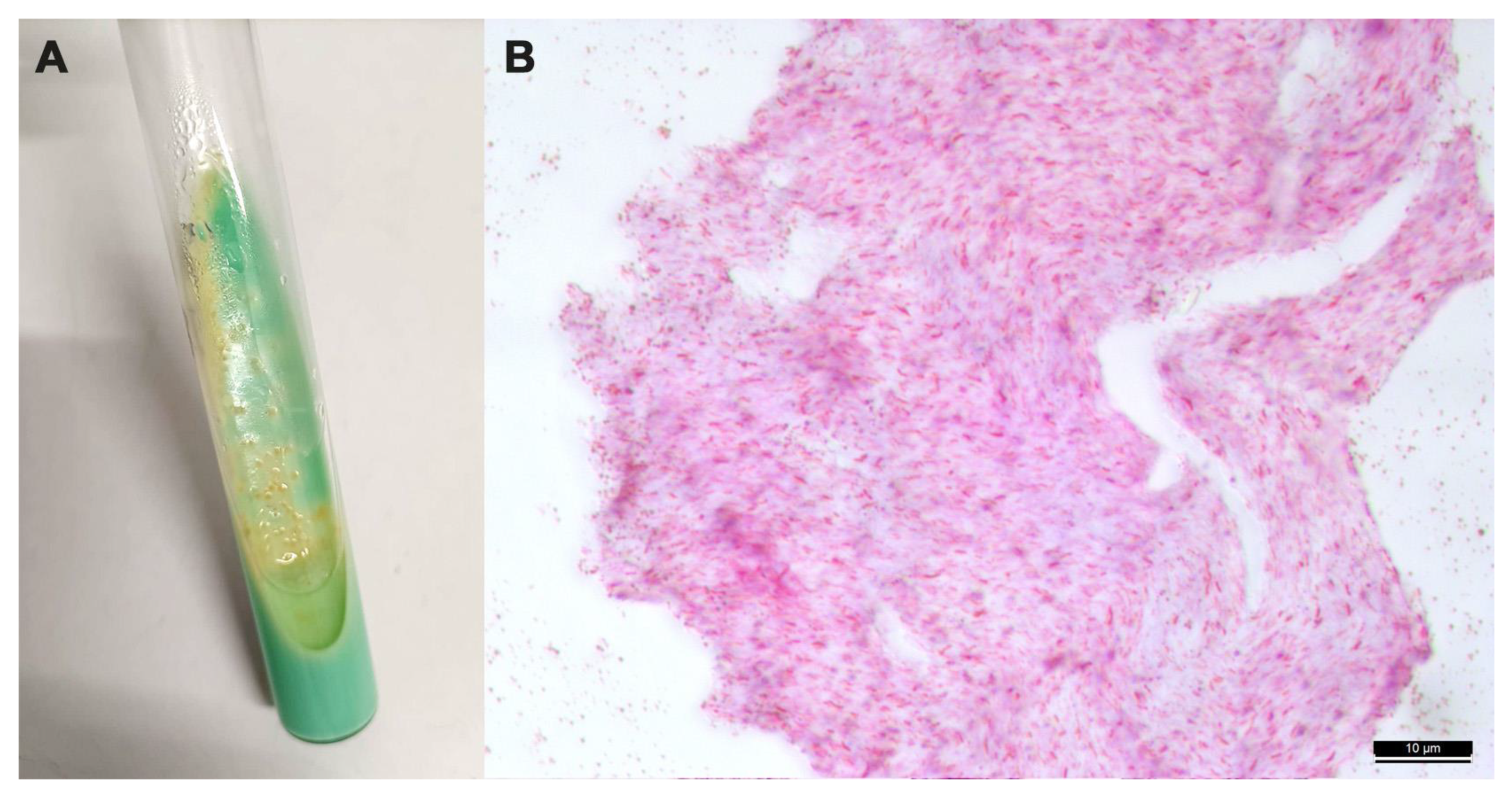
2.3. Bacteriology
2.4. DNA Extraction and IS6110 PCR
2.5. Genotyping of Mycobacterium spp. Isolates
2.6. Statistical Analysis
3. Results
4. Discussion
5. Conclusions
Author Contributions
Funding
Institutional Review Board Statement
Informed Consent Statement
Data Availability Statement
Conflicts of Interest
References
- Tortoli, E.; Fedrizzi, T.; Meehan, C.J.; Trovato, A.; Grottola, A.; Giacobazzi, E.; Serpini, G.F.; Tagliazucchi, S.; Fabio, A.; Bettua, C.; et al. The new phylogeny of the genus Mycobacterium: The old and the news. Infect. Genet. Evol. 2017, 56, 19–25. [Google Scholar] [CrossRef] [PubMed]
- van Ingen, J.; Turenne, C.Y.; Tortoli, E.; Wallace, R.J.; Brown-Elliott, B.A. A definition of the Mycobacterium avium complex for taxonomical and clinical purposes, a review. Int. J. Syst. Evol. Microbiol. 2018, 68, 3666–3677. [Google Scholar] [CrossRef] [PubMed]
- Gortázar, C.; Ferroglio, E.; Höfle, U.; Frölich, K.; Vicente, J. Diseases shared between wildlife and livestock: A European perspective. Eur. J. Wildl. Res. 2007, 53, 241–256. [Google Scholar] [CrossRef]
- Reis, A.C.; Ramos, B.; Pereira, A.C.; Cunha, M.V. The hard numbers of tuberculosis epidemiology in wildlife: A meta-regression and systematic review. Transbound. Emerg. Dis. 2021, 68, 3257–3276. [Google Scholar] [CrossRef]
- Bourne, F.J.; Donnelly, C.A.; Cox, D.R.; Gettinby, G.; McInerney, J.P.; Morrison, W.I.; Woodroffe, R. The Scientific Evidence, a Science Base for a Sustainable Policy to Control TB in Cattle, an Epidemiological Investigation into Bovine Tuberculosis; Final Report of the Independent Scientific Group on Cattle TB; DEFRA: London, UK, 2007; Available online: www.bovinetb.info/docs/final_report.pdf (accessed on 3 April 2023).
- Corner, L.A.; O’Meara, D.; Costello, E.; Lesellier, S.; Gormley, E. The distribution of Mycobacterium bovis infection in naturally infected badgers. Vet. J. 2012, 194, 166–172. [Google Scholar] [CrossRef]
- Byrne, A.W.; Sleeman, D.P.; O’Keeffe, J.; Davenport, J. The ecology of the European badger (Meles meles) in Ireland: A review. In Biology and Environment: Proceedings of the Royal Irish Academy; Royal Irish Academy: Dublin, Ireland, 2012; pp. 105–132. [Google Scholar]
- Swift, B.M.C.; Barron, E.S.; Christley, R.; Corbetta, D.; Grau-Roma, L.; Jewell, C.; O’Cathail, C.; Mitchell, A.; Phoenix, J.; Prosser, A.; et al. Tuberculosis in badgers where the bovine tuberculosis epidemic is expanding in cattle in England. Sci. Rep. 2021, 11, 20995. [Google Scholar] [CrossRef] [PubMed]
- Byrne, A.W.; Kenny, K.; Fogarty, U.; O’Keeffe, J.J.; More, S.J.; McGrath, G.; Teeling, M.; Martin, S.W.; Dohoo, I.R. Spatial and temporal analyses of metrics of tuberculosis infection in badgers (Meles meles) from the Republic of Ireland: Trends in apparent prevalence. Prev. Vet. Med. 2015, 122, 345–354. [Google Scholar] [CrossRef]
- Réveillaud, É.; Desvaux, S.; Boschiroli, M.L.; Hars, J.; Faure, É.; Fediaevsky, A.; Cavalerie, L.; Chevalier, F.; Jabert, P.; Poliak, S.; et al. Infection of wildlife by Mycobacterium bovis in France assessment through a National Surveillance System, Sylvatub. Front. Vet. Sci. 2018, 5, 262. [Google Scholar] [CrossRef]
- Blanco Vázquez, C.; Barral, T.D.; Romero, B.; Queipo, M.; Merediz, I.; Quirós, P.; Armenteros, J.Á.; Juste, R.; Domínguez, L.; Domínguez, M.; et al. spatial and temporal distribution of Mycobacterium tuberculosis Complex infection in Eurasian badger (Meles meles) and cattle in Asturias, Spain. Animals 2021, 11, 1294. [Google Scholar] [CrossRef]
- Jacquier, M.; Vandel, J.M.; Léger, F. Breaking down population density into different components to better understand its spatial variation. BMC Ecol. Evo. 2021, 21, 82. [Google Scholar] [CrossRef]
- Revilla, E.; Delibes, M.; Travaini, A.; Palomares, F. Physical and population parameters of the Eurasian badgers (Meles meles L.) from Mediterranean Spain. Z. Saugetierkd. 1999, 64, 269–276. [Google Scholar]
- Silva, M.; Rosalino, L.M.; Alcobia, S.; Santos-Reis, M. Sett use, density and breeding phenology of badgers in Mediterranean agro-sylvo-pastoral systems. Animals 2021, 11, 2663. [Google Scholar] [CrossRef] [PubMed]
- Gallagher, J.; Clifton-Hadley, R.S. Tuberculosis in badgers; a review of the disease and its significance for other animals. Res. Vet. Sci. 2000, 69, 203–217, Erratum in Res. Vet. Sci. 2001, 70, 179. [Google Scholar] [CrossRef]
- Balseiro, A.; Merediz, I.; Sevilla, I.A.; García-Castro, C.; Gortázar, C.; Prieto, J.M.; Delahay, R.J. Infection of Eurasian badgers (Meles meles) with Mycobacterium avium complex (MAC) bacteria. Vet. J. 2011, 188, 231–233. [Google Scholar] [CrossRef]
- Sistema Informativo Veterinario–Statistiche; Banca Dati Nazionale. Available online: https://www.vetinfo.it/j6_statistiche/#/ (accessed on 23 August 2023).
- Pigozzi, G.; De Marinis, A.M. Meles meles (Linnaeus, 1758). In Fauna d’Italia. Mammalia. III. Carnivora–Artiodactyla; Boitani, L., Lovari, S., Vigna Taglianti, A., Eds.; Calderini: Bologna, Italy, 2003; pp. 158–167. [Google Scholar]
- Johnson, D.D.P.; Macdonald, D.W.; Dickman, A.J. Analysis and review of models of the sociobiology of the Mustelidae. Mammal. Rev. 2000, 30, 171–196. [Google Scholar] [CrossRef]
- Balestrieri, A.; Cardarelli, E.; Pandini, M.; Remonti, L.; Saino, N.; Prigioni, C. Spatial organisation of European badger (Meles meles) in Northern Italy as assessed by camera-trapping. Eur. J. Wildl. Res. 2016, 62, 219–226. [Google Scholar] [CrossRef]
- Regione Molise. Piano Forestale Regionale. Studio di Incidenza Ambientale. 2017. Available online: https://www.regione.molise.it/flex/cm/pages/ServeAttachment.php/L/IT/D/8%252F8%252F6%252FD.d08fdf775275ec8fb3a7/P/BLOB%3AID%3D327/E/pdf?mode=download (accessed on 23 August 2023).
- Pirone, G.; Frattaroli, A.R.; Ciaschetti, G.; Di Martino, L. La vegetazione d’Abruzzo. In Abruzzo Rapporto Sullo Stato Dell’Ambiente 2018; Carsa: Pescara, Italy, 2018; pp. 159–200. Available online: https://www.artaabruzzo.it/download/pubblicazioni/relaz_stato_ambiente_abruzzo_2018.pdf (accessed on 23 August 2023).
- Bern Convention. Convention on the Conservation of European Wildlife and Natural Habitats. Treaty No. 104. Convention on the Conservation of European Wildlife and Natural Habitats. Council of Europe, 1979. Available online: www.coe.int/en/web/bern-convention/ (accessed on 2 April 2023).
- National Law (Italy) n. 503, 05/08/1981. Ratifica ed Esecuzione della Convenzione Relativa alla Conservazione Della vita Selvatica e Dell’ambiente Naturale in Europa, Con Allegati, Adottata a Berna il 19 Settembre 1979. Off. J. 250, 11/09/1981. Available online: https://www.normattiva.it/uri-res/N2Ls?urn:nir:stato:legge:1981-08-05;503 (accessed on 23 August 2023).
- Anderson, R.M.; Trewhella, W. Population dynamics of the badger (Meles meles) and the epidemiology of bovine tuberculosis (Mycobacterium bovis). Philos. Trans. R. Soc. Lond. B Biol. Sci. 1985, 310, 327–381. [Google Scholar]
- Bertusi, M.G.; Tosetti, T. Tasso (Meles meles). In Mammiferi d’Italia; Grafis: Bologna, Italy, 1986. [Google Scholar]
- Delahay, R.J. A forensic investigation of Mycobacterium bovis infection in badgers. Vet. J. 2012, 194, 135–136. [Google Scholar] [CrossRef]
- World Organization for Animal Health. Bovine Tuberculosis. In Manual of Diagnostic Tests and Vaccines for Terrestrial Animals; OIE: Paris, France, 2018; pp. 1058–1074. [Google Scholar]
- Boniotti, M.B.; Gaffuri, A.; Gelmetti, D.; Tagliabue, S.; Chiari, M.; Mangeli, A.; Spisani, M.; Nassuato, C.; Gibelli, L.; Sacchi, C.; et al. Detection and molecular characterization of Mycobacterium microti isolates in wild boar from Northern Italy. J. Clin. Microbiol. 2014, 52, 2834–2843. [Google Scholar] [CrossRef]
- Collins, D.M.; Stephens, D.M.; de Lisle, G.W. Comparison of polymerase chain reaction tests and faecal culture for detecting Mycobacterium paratuberculosis in bovine faeces. Vet. Microbiol. 1993, 36, 289–299. [Google Scholar] [CrossRef]
- Kulski, J.K.; Khinsoe, C.; Pryce, T.; Christiansen, K. Use of a multiplex PCR to detect and identify Mycobacterium avium and M. intracellular in blood culture fluids of AIDS patients. J. Clin. Microbiol. 1995, 33, 668–674. [Google Scholar] [CrossRef] [PubMed]
- Wilton, S.; Cousins, D. Detection and identification of multiple mycobacterial pathogens by DNA amplification in a single tube. PCR Methods Appl. 1992, 1, 269–273. [Google Scholar] [CrossRef] [PubMed]
- Sivia, D.S. Data Analysis: A Bayesian Tutorial; Oxford University Press: Oxford, UK, 1996. [Google Scholar]
- Fabrizio, M.; Di Febbraro, M.; D’Amico, M.; Frate, L.; Roscioni, F.; Loy, A. Habitat suitability vs. landscape connectivity determining roadkill risk at a regional scale: A case study on European badger (Meles meles). Eur. J. Wildl. Res. 2019, 65, 7. [Google Scholar] [CrossRef]
- Glawischnig, W.; Steineck, T.; Spergser, J. Infections caused by Mycobacterium avium subspecies avium, hominissuis, and paratuberculosis in free-ranging red deer (Cervus elaphus hippelaphus) in Austria, 2001–2004. J. Wildl. Dis. 2006, 42, 724–731. [Google Scholar] [CrossRef] [PubMed]
- Carta, T.; Álvarez, J.; Pérez de la Lastra, J.M.; Gortázar, C. Wildlife and paratuberculosis: A review. Res. Vet. Sci. 2013, 94, 191–197. [Google Scholar] [CrossRef]
- Whittington, R.; Donat, K.; Weber, M.F.; Kelton, D.; Nielsen, S.S.; Eisenberg, S.; Arrigoni, N.; Juste, R.; Sáez, J.L.; Dhand, N.; et al. Control of paratuberculosis: Who, why and how. A review of 48 countries. BMC Vet. Res. 2019, 15, 198. [Google Scholar] [CrossRef]
- Attili, A.R.; Ngu Ngwa, V.; Preziuso, S.; Pacifici, L.; Domesi, A.; Cuteri, V. Ovine paratuberculosis: A seroprevalence study in dairy flocks reared in the Marche region, Italy. Vet. Med. Int. 2011, 2011, 782875. [Google Scholar]
- Iarussi, F.; Paradies, P.; Sardaro, R.; Rubino, G.; Scaltrito, D.; Pieragostini, E.; Petazzi, F. Epidemiology and risk factors of Mycobacterium avium subspecies paratuberculosis in semi-extensive dairy sheep and goat farms of Apulia, Southern Italy. Small Rumin. Res. 2019, 177, 89–96. [Google Scholar] [CrossRef]
- Cenci-Goga, B.T.; Vescera, F.; Paolotto, P.; McCrindle, C.M.; Roberti, U. Seroprevalence of Mycobacterium avium subspecies paratuberculosis in cows in Umbria, Italy. Vet. Rec. 2010, 167, 577–578. [Google Scholar] [CrossRef]
- Marchetti, G.; Ricchi, M.; Serraino, A.; Giacometti, F.; Bonfante, E.; Arrigoni, N. Prevalence of Mycobacterium avium subsp. paratuberculosis in milk and dairy cattle in Southern Italy: Preliminary results. Ital. J. Food Saf. 2013, 2, e35. Available online: https://www.pagepressjournals.org/index.php/ijfs/article/view/ijfs.2013.e35 (accessed on 23 August 2023).
- Fanelli, A.; Buonavoglia, D.; Pleite, C.M.C.; Tizzani, P. Paratuberculosis at European scale: An overview from 2010 to 2017. Vet. Ital. 2020, 56, 13–21. [Google Scholar]
- Zanetti, S.; Bua, A.; Molicotti, P.; Delogu, G.; Mura, A.; Ortu, S.; Sechi, L.A. Identification of mycobacterial infections in wild boars in Northern Sardinia, Italy. Acta Vet. Hung. 2008, 56, 145–152. [Google Scholar] [CrossRef] [PubMed]
- Barry, C.; Corbett, D.; Bakker, D.; Andersen, P.; McNair, J.; Strain, S. The effect of Mycobacterium avium Complex infections on routine Mycobacterium bovis diagnostic tests. Vet. Med. Int. 2011, 2011, 145092. [Google Scholar] [CrossRef] [PubMed]
- Byrne, A.W.; Graham, J.; Milne, G.; Guelbenzu-Gonzalo, M.; Strain, S. Is there a relationship between bovine Tuberculosis (bTB) herd breakdown risk and Mycobacterium avium subsp. paratuberculosis status? An investigation in bTB chronically and non-chronically infected herds. Front. Vet. Sci. 2019, 6, 30. [Google Scholar] [PubMed]
- Picasso-Risso, C.; Grau, A.; Bakker, D.; Nacar, J.; Mínguez, O.; Perez, A.; Alvarez, J. Association between results of diagnostic tests for bovine tuberculosis and Johne’s disease in cattle. Vet. Rec. 2019, 185, 693. [Google Scholar] [CrossRef]
- Stabel, J.R.; Waters, W.R.; Bannantine, J.P.; Palmer, M.V. Comparative cellular immune responses in calves after infection with Mycobacterium avium subsp. paratuberculosis, M. avium subsp. avium, M. kansasii and M. bovis. Vet. Immunol. Immunopathol. 2021, 237, 110268. [Google Scholar] [CrossRef]
- Galiero, A.; Leo, S.; Garbarino, C.; Arrigoni, N.; Russo, S.; Giacomelli, S.; Bianchi, A.; Trevisiol, K.; Idrizi, I.; Daka, G.; et al. Mycobacterium aviumsubsp. paratuberculosis isolated from wild red deer (Cervus elaphus) in Northern Italy. Vet Microbiol. 2018, 217, 167–172. [Google Scholar] [CrossRef]
- Fico, R.; Mariacher, A.; Franco, A.; Eleni, C.; Ciarrocca, E.; Pacciarini, M.L.; Battisti, A. Systemic tuberculosis by MYCOBACTERIUM BOVIS in a free-ranging MARSICAN brown bear (URSUS ARCTOS MARSICANUS): A Case report. BMC Vet. Res. 2019, 15, 152. [Google Scholar] [CrossRef]
- European Regulation EU 2021/620 Commission Implementing Regulation (EU) 2021/620 of 15 April 2021 Laying Down Rules for the Application of Regulation (EU) 2016/429 of the European Parliament and of the Council as Regards the Approval of the Disease-Free and Non-Vaccination Status of Certain Member States or Zones or Compartments Thereof as Regards Certain Listed Diseases and the Approval of Eradication Programmes for Those Listed Diseases. Available online: https://eur-lex.europa.eu/legal-content/EN/TXT/?uri=CELEX%3A02021R0620-20231127 (accessed on 21 December 2023).
- Abbate, J.M.; Arfuso, F.; Iaria, C.; Arestia, G.; Lanteri, G. Prevalence of Bovine Tuberculosis in Slaughtered Cattle in Sicily, Southern Italy. Animals 2020, 10, 1473. [Google Scholar] [CrossRef]
- Di Marco, V.; Mazzone, P.; Capucchio, M.T.; Boniotti, M.B.; Aronica, V.; Russo, M.; Fiasconaro, M.; Cifani, N.; Corneli, S.; Biasibetti, E.; et al. Epidemiological significance of the domestic black pig (Sus scrofa) in maintenance of bovine tuberculosis in Sicily. J. Clin. Microbiol. 2012, 50, 1209–1218. [Google Scholar] [CrossRef]
- Amato, B.; Di Marco Lo Presti, V.; Gerace, E.; Capucchio, M.T.; Vitale, M.; Zanghì, P.; Pacciarini, M.L.; Marianelli, C.; Boniotti, M.B. Molecular epidemiology of Mycobacterium tuberculosis complex strains isolated from livestock and wild animals in Italy suggests the need for a different eradication strategy for bovine tuberculosis. Transbound. Emerg. Dis. 2018, 65, e416–e424. [Google Scholar] [CrossRef] [PubMed]


| Mycobacterium Target | Name of the Primer | Primer Sequencing | Target Gene | Amplicon Size |
|---|---|---|---|---|
| Mycobacterium spp. | Mycgen-F | 5′-AGAGTTTGATCCTGGCTCAG-3′ | mbp70 | 1030 bp |
| Mycgen-R | 5′-TGCACACAGGCCACAAGGGA-3′ | |||
| M. tuberculosis complex | TB1-F | 5′-GAACAATCCGGAGTTGACAA-3′ | mbp70 | 372 bp |
| TB1-R | 5′-AGCACGCTGTCAATCATGTA-3′ | |||
| M. avium paratuberculosis | DMC1 | 5′-GATCGGAACGTCGGCTGGTCAGG-3′ | IS900 | 217 bp |
| DMC2 | 5′-GATCGCCTTGCTCATCGCTGCCG-3′ | |||
| M. avium avium | Mycgen-F | 5′-AGAGTTTGATCCTGGCTCAG-3′ | mbp70 | 180 bp |
| Mycav-R | 5′-ACCAGAAGACATGCGTCTTG-3′ |
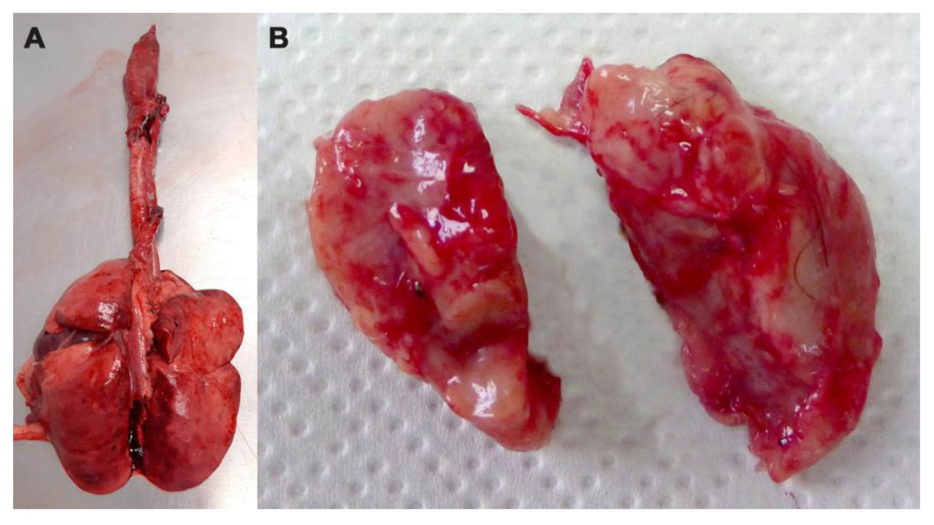
| General Register Number | Date of Discovery | Municipality of Discovery | Sex | Age | Cause of Death | Tissues Examined | Methods | Mycobacterium Species |
|---|---|---|---|---|---|---|---|---|
| 4860AZ2017 | 11 September 2017 | Introdacqua (AQ) | F | Adult | Road-killed | Head lymph nodes | Culture+; BACTEC- PCR- | M. avium subsp. avium |
| 4804AZ2019 | 4 September 2019 | Collelongo (AQ) | F | Adult | Road-killed | Head lymph nodes | Culture+; BACTEC+ PCR- | M. spp. |
| 41362TE2020 | 7 May 2020 | Ortona (CH) | M | Adult | Predation | Head lymph nodes | Culture+; BACTEC+ PCR- | M. avium subsp. paratuberculosis |
| 298930TE2020 | 26 November 2020 | Vacri (CH) | M | Young | Road-killed | Head lymph nodes | Culture+; BACTEC+ PCR- | M. avium subsp. avium |
| Mediastinal lymph nodes | Culture-; BACTEC-; PCR- | |||||||
| Lung | Histological exam: absence of tubercular lesion |
Disclaimer/Publisher’s Note: The statements, opinions and data contained in all publications are solely those of the individual author(s) and contributor(s) and not of MDPI and/or the editor(s). MDPI and/or the editor(s) disclaim responsibility for any injury to people or property resulting from any ideas, methods, instructions or products referred to in the content. |
© 2024 by the authors. Licensee MDPI, Basel, Switzerland. This article is an open access article distributed under the terms and conditions of the Creative Commons Attribution (CC BY) license (https://creativecommons.org/licenses/by/4.0/).
Share and Cite
Tieri, E.E.; Marino, L.; Zilli, K.; Pompilii, C.; Di Teodoro, G.; Cocco, A.; Ruberto, A.; Toro, M.; Mastrodomenico, M.T.; Salucci, S.; et al. Survey of Mycobacterium spp. in Eurasian Badgers (Meles meles) in Central Italy. Animals 2024, 14, 219. https://doi.org/10.3390/ani14020219
Tieri EE, Marino L, Zilli K, Pompilii C, Di Teodoro G, Cocco A, Ruberto A, Toro M, Mastrodomenico MT, Salucci S, et al. Survey of Mycobacterium spp. in Eurasian Badgers (Meles meles) in Central Italy. Animals. 2024; 14(2):219. https://doi.org/10.3390/ani14020219
Chicago/Turabian StyleTieri, Elga Ersilia, Lucio Marino, Katiuscia Zilli, Cinzia Pompilii, Giovanni Di Teodoro, Antonio Cocco, Addolorato Ruberto, Michela Toro, Maria Teresa Mastrodomenico, Stefania Salucci, and et al. 2024. "Survey of Mycobacterium spp. in Eurasian Badgers (Meles meles) in Central Italy" Animals 14, no. 2: 219. https://doi.org/10.3390/ani14020219